


![]()
![]()
![]()
BibTeX | RIS | EndNote | Medlars | ProCite | Reference Manager | RefWorks
Send citation to:
URL: http://ijmm.ir/article-1-1805-en.html


2- Department of Pathobiology, Faculty of Veterinary Medicine, Lorestan University, Khorramabad, Iran ,
3- Department of Pathobiology, Faculty of Veterinary Medicine, Semnan University, Semnan, Iran
Leptospira are one of the most widespread bacteria which can infect both humans and animals (1). These bacteria are not the microbiota of the human and animal bodies. They are aquatic and mobile objects which live naturally in humid areas such as moist soil, freshwater, sludge, and vegetation for long periods. In humans, direct contact with infected animals is the most known transmission route of leptospira. Moreover, animals are usually contaminated via soil, water, and body fluids which constantly excrete bacteria through urine, genital secretions, milk, and infected aerosols. It has been reported these bacteria in infected and asymptomatic animals are able to survive for a couple of months in the proximal tube of the kidney and are excreted in the urine, consequently, it can increase the contamination rate of the environment and transmit the disease to human or other animals (1-3).
Leptospirosis is currently known as one of the major health problems in the world. The commonality of this disease between humans and animals can highlight the importance of this disease (4). This disease not only can extremely threaten public health but also causes major economic losses to the country's livestock industry (e.g., abortion, poor births, fertility…). It must be mentioned that abortion is considered the most prevalent symptom of leptospirosis in sheep (2, 4, 5).
Since sheep husbandry plays a vital role in the livestock industry of many zones of Iran, such as Lorestan province, hence abortion is able to directly impress the economy of these provinces. Given the issue's importance in public health and economic losses, planning and implementing scientific standards in line with the importance of abortion management factors can be very influential in reducing this issue and preventing the resulting economic losses. Therefore, the aim of this study was to investigate the presence of Leptosoira spp. genomic DNA using PCR in the vaginal swab samples of sheep with abortion history in Lorestan province, Iran.
Sample Collection
In this cross-sectional descriptive study, a total of 150 samples of vaginal swabs were collected from different areas of Lorestan province. It is important to note that the abortion history of the sheep which were selected for sampling, was less than a month. Moreover, the sampling was randomly performed with the cooperation of veterinary offices of Khorramabad, Chegni, Boroujerd, Noorabad, Kuhdasht, and Poldakhtar cities during the spring and autumn seasons from May 2019 to June 2020. The collected samples were transferred to tubes containing phosphate-buffered saline (PBS) and stored at -20°C until performing the project.
DNA extraction and amplification
To extract Leptosoira spp. genomic DNA of the collected samples, the DNA extraction kit (GeneAll® Exgene™ Cell SVmini-250, Korea), was employed. The DNA extraction procedure was performed based on the manufacturer's instructions. PCR as a robust molecular tool was applied to amplify Leptosoira spp. DNA in the collected samples. Nucleotide sequences were selected for lipL32 gene amplification. The amplification was conducted in 25 µL total volume using 12.5 μL of 2X master mix (Ampliqon Taq DNA Polymerase Master Mix RED), 0.5 μL of each specific forward and reverse primers (10nM) (provided by Takapou Zist Company, Tehran, Iran), and 5 μL of the extracted DNA (Table 1).
The current study used standard L. interrogans genomic DNA donated by the L. interrogans Research Laboratory of the University of Tehran as a positive control (L. interrogans ATCC 43642). Also, all PCR reagents except extracted DNA were mixed and applied to prepare negative control. The PCR amplifications were performed by Bio-Rad thermocycler (Model T- 100, USA) under the following conditions: The initial step of 94°C for 2 min, followed by 35 cycles of 94°C for 30 s as denaturation, annealing at 59°C for 30 Second, extension at 72°C for 1 min, and followed by a final extension at 72°C for 5 min.
To investigate the PCR products, a 1.5% agarose gel (Merck, Germany) containing 2 μg/mL DNA-safe stain (Cinnagen, Iran) was used. After electrophoresis, the gel was placed in a Trans illuminator (E-Box, Iran) to visualize the specific bands. Finally, the PCR products with 242bp in length were compared with ladder and positive /negative controls.
Table 1. The sequence of specific primers used to amplify the genotype of Leptosoira spp.
| Reference | Amplification Size (bp) | Primer sequence | Primer name | Target gene |
| (6) | 242 | 5-AAG CAT TAC CGC TTG TGG TG-3 | F1 | Lip L 32-45F |
| 5-GAA CTC CCA TTT CAG CGA TT-3 | R1 | Lip L 32-286R |
The results of this study revealed that out of 150 samples of vaginal swabs collected from sheep with an abortion history in different areas of Lorestan province, generally, two samples (1.3%) were diagnosed as positive cases by PCR. All positive samples of the 242bp band were observed (Figure 1). The results of data analysis demonstrated that among the different areas which was selected for sampling pollution was traced only to Khorramabad city and the rest of the cities had no infection (Table 2).
Leptospirosis is one of the most important diseases with global spread which leads to significant economic damage to the country's livestock industry through the increase in mortality rate, a decrease in milk production, and an increase in reproductive disorders(e.g. abortion) (7, 8). Many records from the scientific centers demonstrated that this infection is one of the problems of the livestock industry in most parts of Iran. Due to the characteristics of this bacterium, it can properly survive in wet soil and water, therefore this bacterium is mainly found in areas with hot and humid climates and heavy rainfall, such as the northern provinces and Khuzestan (9-12). Since no study has been conducted on the prevalence of this disease in sheep in Lorestan province, the current study was designed to investigate the prevalence rate of this bacterium in sheep with an abortion history.
Table 2. Frequency and percentage of positive cases of Leptospira infection in vaginal swab samples of aborted sheep in Lorestan province
| Number of negative cases | Number of positive Cases | Number of Samples | Sampling Season | Sampling Location |
| 87 (7/97 %) | 2 (%2.3) | 89 | Spring | Khorramabad and Dore Chegni |
| 3 (100) | (0)0 | 3 | Fall | Borujerd |
| 11 (100) | (0)0 | 11 | Fall | Koohdasht |
| 17 (100) | (0)0 | 17 | Fall | Noor-Abad |
| 30 (100) | (0)0 | 30 | Fall | Poldokhtar |
| 148 (98.7%) | 1.3 % | 150 | Total | |
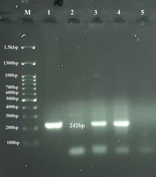
Figure 1. The PCR results of samples on agarose gel 1.5%. M; 100 bp DNA Ladder; Column 1: positive control; column 2: negative control; columns 3,4: positive samples; columns 5: negative sample. The desired band at this stage is 242bp
Due to technical problems of culture and isolation of the causative agent of this disease, such as time-consuming and low sensitivity, the major studies which are designed for the diagnosis of this disease are based on serological and molecular methods (13). Currently, serological tests are taken into account as the most common diagnostic test for the detection of this bacterium. MAT is the most important standard serological test used to detect anti-Leptospira antibodies in the serum. However, there are several limitations to applying this test, including the necessity of maintaining a wide range of Leptospira serovars to provide a live antigen source, the need for standard antiserums and dark field microscope, the skill and expertise of laboratory technicians to have paired serum samples that delay diagnosis, and false-negative results, which are mainly due to not testing with all serotypes. However, interpretation of MAT is difficult due to the high rate of cross-reactions that occur among different serogroups, especially in the acute phase. Due to the mentioned points and limitations in the diagnosis of chronic infections with the MAT method, it seems that molecular methods such as PCR can be reliably, and rapidly performed on a wide variety of samples such as urine, milk, liver, kidney, and vaginal secretions (14, 15). In this study, the PCR method was used to detect the presence of lipL32 gene, which encodes one of the most important immunogenic proteins of the Leptospira spp. outer membrane. In this study, which was analyzed out of samples of vaginal swabs by the PCR method 1.3% of samples were positive for Leptospira spp. The results of this study were confirmed with the results of a study performed by Haji Hajikolaei et al. (11). The results showed a low percentage of infection in sheep in the region. According to the results of their study, the positive cases of the disease were related to the season (spring, which may be due to the favorable weather conditions and rainfall) and applying a traditional feeding system. Considering the mountainous climate of Lorestan province and considering that most of the studied areas are non-swampy environments and belong to cold and mountainous climates, the type of sample collection, the applied technique, and the low percentage of positive samples can be justified. Based on the studies performed, the best sample to diagnose abortion because of leptospira is sampling from the abomasum of an aborted fetus, and if the fetus is not available vaginal sampling is important (16-18). In view of this point and the density of livestock population in the province, the traditional method of husbandry and raising sheep, and the climate of Lorestan province, it seems that the percentage obtained in this study cannot accurately justify the pollution situation in this province. Babakhani et al. (10) in a study of a total of 200 serum samples taken from Wisian paddy farmers in Lorestan province by MAT method, reported a 30% prevalence of serum contamination with Leptospira serovars. The most important reason for this difference can be related to the type of technique used and the selection of the sample under study.
Hamli et al. (12), in the study of leptospira-induced abortion in cows of Chaharmahal Bakhtiari province by PCR method, from 120 samples collected from the abomasum of an aborted fetus, reported 17 positive samples (14.16%) (12). The most important reason for the difference between the results of Hamlin's study and the current study is probably related to the type of sample studied and the animal species. According to sources, in cattle, unlike sheep, bacteria except in the liver, kidneys, lungs, placenta, and uterus of the cow can colonize in the vaginal secretions of cows. In the case of sheep, this bacteria is mainly located in the kidneys and can cause pyelonephritis, consequently, it is widely spread through urine in the surrounding environment (16, 17). Almeida et al. (3) sampled vaginal secretions from 15 ewes of a herd in Brazil that had no history of abortion or reproductive disorders, and their serum contamination had previously been confirmed by MAT. In the mentioned study, after PCR using LipL32 gene primers, seven samples (46.7%) contained the Leptospira genome. Because Leptospira is mainly located in the kidneys of sheep and subsequently the bacteria can spread through urine in the environment, it is possible that the found DNA of the Leptospira in the present study and other researchers, such as Almeida et al. (3) contains bacteria due to vaginal contamination of urine. The results of this study and another study by researchers (16-18) indicate that whether the vagina is primarily infected by bacteria or secondarily by urine passage, there is a possibility of disease transmission from female animals and the potential for infection from male sheep through sexual transmission. However, since Leptospira is traceable in semen and sexual secretions of male animals and sexual transmission from male sheep to ewes is well identified, this may be the reason for the infection of female sheep in this study.
A brief look at the mentioned studies shows that there are differences between various regions in terms of serum and molecular contamination with Leptospira. These differences can be related to geographical conditions, animal species, amount of stagnant and surface water in the area, location and method of livestock (traditional or industrial), quality of farms and the number of livestock, simultaneous maintenance of several types of livestock, herd health, vaccination, ambient temperature, and annual rainfall.
Unfortunately, few studies have been conducted on the small role of this bacterium in sheep abortion, it seems that the frequency of this crime in sheep herds in Lorestan province is more than the amount obtained in this study. Due to the zoonotic nature of this bacterium and the importance of diagnosing the disease in livestock to control the disease in humans, rapid diagnostic methods such as PCR and using serological tests with a sampling of the abomasum of an aborted fetus are used considered necessary.
The authors thank the staff of veterinary offices in Khorramabad, Boroujerd, Noorabad, Poldakhtar, and Kuhdasht for helping to collect samples.
This research is financially supported by Lorestan University, Iran.
Conflicts of Interest
The authors declare that they have no conflict of interest.
Received: 2022/07/4 | Accepted: 2022/09/11 | ePublished: 2023/01/20
| Rights and permissions | |
 |
This work is licensed under a Creative Commons Attribution-NonCommercial 4.0 International License. |
Copyright Policy
Iranian Journal of Medical Microbiology by Farname is licensed under CC BY-NC 4.0![]()
![]()
![]()
