


![]()
![]()
![]()
BibTeX | RIS | EndNote | Medlars | ProCite | Reference Manager | RefWorks
Send citation to:
URL: http://ijmm.ir/article-1-1074-en.html


2- Farzanegan 2 Highschool, Tehran, Iran
.
Licorice with the scientific name of Glycyrrhiza glabra is a plant native to the Mediterranean region and grows in most parts of Iran. This perennial herbaceous plant has numerous underground stems and its leaves are compound. Its flowers are bluish and its fruits contain 5 to 6 rosary-shaped and brown seeds. This plant belongs to the butterfly family and has been one of the most important native medicinal plants in Iran since ancient times, which grows in most regions of Iran. Two varieties named glabra and glandulifera (Waldst & Kit) Boiss have been identified and the most important collection areas are Fars province. It is widely used in traditional medicine and treatment of digestive problems (1). G. glabra rhizomes contain many active ingredients including triterpene, saponins, glycyrrhizin and 2,4-hydroxyglycyrrhizin, which have a sweetening power of 100-50 times higher than sugar cane. Flavonoids such as isoliquiritigenin liquiritigenin, isolicoflavonol, and isoflavones such as glabren, glabridin, glabrol, 3-hydroxyglabroI, and glycyrrhisoflavone are also abundant in this plant root. Other compounds, including hydroxycoumarin include herniarin umbelliferone, glycycoumarin, licopyranocumarin and cumin derivatives, including glycerol, isoglycyrol, liqcoumarin and steroids, including stereos, beta-sterols stigmasterol and fructose oils including anethole, Licorice is found. Licorice has a mild nature in terms of ancient Iranian medicine. The most important newly discovered property of licorice, used in Germany, Europe and the United States, is to treat stomach ulcers and stomach cancer. Licorice rhizome and its components can have beneficial effects in the prevention and treatment of gastrointestinal diseases, especially gastric ulcer, bloating, constipation, tonsillitis and hepatitis. On the other hand, the effects of licorice and its active components on the respiratory system and elimination of cough, asthma, and chest infections have been observed in studies (2).
Today, the anti-cancer effects of some Glycyrrhiza glabra compounds have been identified. Also, the beneficial effects of licorice on the treatment of depression indicate a wide range of capabilities of this plant. Licorice and its active ingredients have a wide range of beneficial antioxidant, anti-inflammatory and anti-allergic effects. Therefore, in Phytotherapy, special attention should be paid to this plant. (5, 6 and25) According to the articles, excessive consumption of licorice causes high blood pressure, so it is better to consume tragacanth to eliminate the blood pressure caused by eating licorice plants to eliminate this complication (7). Khoshnam et al. In 2016 proved in a study that the combination of licorice (90 mg/ kg) with LNAME has synergistic effects on lowering blood pressure (8).
Tragacanth with the scientific name of Astraglus gossypinus, is the gum and sap of a plant called Astragalus, which has different species in the form of shrubs or perennial and annual herbaceous plants. This plant has more than 200 species that are mostly grown in steppe and mountainous areas of Iran.
Tragacanth also has a warmer nature. This gum flows spontaneously or usually due to a crack in the stem and dries after a short time, which is collected and marketed in two main forms. The start of operation is usually from late spring to early September. Operators first empty around the stem and then make incisions parallel to the stem (parallel to the phloem) with special blades.
After a few days, the sap (tragacanth) seeps out due to an incision made in the stem, and the sap loses its water and hardens when exposed to sunlight. Operators collect tragacanth twenty days after cutting and repeat the process. In the past, people used tragacanth to wash dry hair. This compound is used to strengthen normal and oily hair and to strengthen dry hair, cough and sputum. Tragacanth contains 10-15% water, 3-4% minerals and 3% starch, and the molecular weight of tragacanth is over eight hundred thousand daltons. Tragacanth is an odorless substance that does not dissolve 60-70% in water, but by absorbing moisture, it becomes a sticky glaze in which starch particles can be seen.
In tragacanth there are generally two types of significant active substances, one is a substance that is soluble in water called tragacanthine, and the second is a substance that is insoluble in cold water and is called basorin. Tragacanthine is composed of galacturonic acid, which is attached to the sugars galactose and xylose. Basurin also contains galacturonic acid bound to the sugars galactose and xylose. These effective substances prevent the prolongation of the stoppage time of feces in the intestine and ultimately prevent the adverse effects of toxins on intestinal tissue and thus prevent the carcinogenic effects of fecal matter (7). Due to the nativeness, I was cheap and the availability of this herb, these plants can be a good alternative to chemical drugs effective in the treatment of gastric ulcer and have the least side effects after consumption.
Gastric or peptic ulcer is one of the most important diseases in which many people in the community are affected. Gastric or peptic ulcer: It is a painful lesion that occurs on the inner wall of the stomach or at the beginning of the small intestine called the duodenum. No known single cause of gastric ulcer has been found. However, it is now clear that gastric ulcer is the result of an imbalance of gastrointestinal and duodenal fluids. One of the causes of stomach ulcers is an infection caused by a bacterium called Helicobacter pylori. Also, the use of painkillers called nonsteroidal anti-inflammatory drugs or NSAIDS, including aspirin, naproxen, ibuprofen and many other drugs can also be effective in treating the symptoms of gastric ulcer (3). Family history of gastric ulcer, other diseases such as liver, kidney or lung diseases, frequent drinking of alcohol and over 50 years of age are also effective in this disease (7).
Some symptoms of stomach ulcers include burning pain in the middle or upper part of the stomach between meals or at night, bloating, heartburn, nausea or vomiting, black or dark stools (due to bleeding), high blood pressure, weight loss, and severe pain. It is in the middle or upper part of the stomach (7).
Half of the people in the community suffer from stomach problems and do not have a proper treatment plan. There are methods for treating stomach ulcers such as antibiotics, endoscopy and surgery. Drugs such as Prilosec, Prevacid, Aciphex, Protonix, Zegerid are used in the treatment of gastric ulcers, which have many side effects and are expensive (9).
Providing a suitable herbal medicine can be a good alternative to chemical treatments for this disease and this is very important. In addition, herbal medicines have the least side effects compared to chemical medicines. The use of native medicinal plants can be the cheapest and available to the patient and reduce the cost of importing medicinal plants and chemical drugs.
This study is related to a two-person experimental study of seventh grade students of Farzanegan Two High School under the guidance of Ms. Bozorgzadeh, High School Research Secretary and Dr. Larypoor, an Assistant Professor of the faculty of Islamic Azad University, North Tehran Branch is done.
Evaluation of Effective Doses of Glycyrrhiza glabra
In order to evaluate the anti-ulcer effects of aqueous extract of Glycyrrhiza glabra, rating and teal flick tests were used. Also, Lorek's method was used to evaluate the acute toxicity of the extract (21). The results of these tests show that the aqueous extract of licorice at a dose of 200 mg/kg clearly showed the anti-ulcer effect on the writing and tealflick tests. Dad. (P<0.01)
Rating Test
In this experiment, adult male NMRI mice weighing 25-35 g were used and on the day of the experiment, in order to accustom the animals to the environment, each of them was placed in the standard glass box 30 minutes before the start of the experiment. Aqueous extract of Glycyrrhiza glabra was dissolved in sterile saline and injected intraperitoneally at doses of 100, 200 and 300 mg / kg body weight (4, 22). After 15 minutes, acetic acid in a volume of 0.1 ml / kg was injected at a concentration of 0.6%, and after intraperitoneal injection of acetic acid, the number of abdominal contractions was counted for 30 minutes. In addition, each animal was used only once in the control group, after intraperitoneal injection of saline, a rating test was performed (21-23).
Carrageenan test
To perform the carrageenan test, first, adult male NMRI mice weighing 25-35 g in seven groups were treated with the desired substance (saline, extract, dexamethasone) in the amount of 0.2 mL intraperitoneally. One hour later, 0.1 mL of 1% carrageenan was injected subcutaneously into the dorsal surface of the animal's right foot. Four hours after carrageenan injection, mice were killed by chloroform and their legs were amputated. Inflammation index was assessed (24). All data were statistically analyzed using one-way ANOVA and Tukey test. Results were presented as a mean ± standard deviation (S.E.M ± Mean). The criterion for statistical inference is (P<0.05) (24).
Preparation of Medicinal Plant
The root of licorice identified with the number IAUNT17333 was prepared from the herbarium of Islamic Azad University, North Tehran Branch and after cleaning, it was divided into 1 cm pieces and ground. It was then poured onto a cloth and 300 mL of 70% methanol was poured into a Soxhlet balloon. After 24 hours, the solvent was gradually evaporated by heat and the active ingredients were transferred to the solvent. The extract was then filtered and transferred to a rotary apparatus at 45 ° C and 45 ° C for one hour to separate the solvent. The extract was then transferred to a dark container and stored in the refrigerator for subsequent experiments (10, 3).
Edible tragacanth with the number IAUNT17334 was prepared from the herbarium of Islamic Azad University, North Tehran Branch, and after powdering, it was dissolved in sterile distilled water at a temperature of 45°C and used. In this study, based on the rating test, carrageenan and determination of toxicity dose and also based on the results of research of Sepehri et al. (2007), the dose of these two herbal medicines was determined to be 200 mg / kg. (4, 22) All solutions were stored at 4°C for later use.
(LD50) Determination of Acute Toxicity
This test was performed based on the method of Dietrich Lorke (1983) (21). After intraperitoneal injection of dilutions of the extract at doses of 10, 100, 1000, 1600, 2900 and 5000 kg / mg, the mortality rate (LD50) of 50% of mice up to 72 hours after injection was evaluated.
The Effect of Aqueous Extract of Glycyrrhiza glabra and Tragacanth in vivo
31 NMRI rats weighing 200-230 g were obtained from Pasteur Institute of Iran. The animals were kept in cages with long mesh floors to prevent defecation and at a temperature of 22-25°C and a period of light and darkness for12 hours and were fed with normal food and free access to water. In order to empty the stomach, all animals except the negative control were kept hungry for 48 hours and fed with 8% sucrose solution during the starvation period to avoid dehydration. Then 4 groups of mice were gavaged for 3 days with aspirin at a dose of 200 mg dissolved in 1 cc of solvent (1% carboxymethylcellulose) based on daily weight of mice. The absence of halal wound healing effect has already been confirmed (3, 4).
Depending on the type of treatment for 14 days, each group received a veterinary licorice extract (concentration of 0.036 mg/mL per compound) and distilled water at a dose of 200 mg by gavage (10, 19). These mice were classified into five groups and each group had 6 mice and were treated for two weeks. The experimental groups were:
1- The first group of mice with gastric ulcer that were treated with licorice extract of Tragacanth at a dose of 200 mg based on daily weight of mice.
2- The second group of mice with gastric ulcer that were treated with licorice extract at a dose of 200 mg based on daily weight of mice.
3- The third group of mice with gastric ulcer that were treated with omeprazole at a dose of 200 mg based on daily weight of mice.
4- The fourth group or positive control of mice with gastric ulcer that were treated with distilled water at a dose of 200 mg based on daily weight of mice.
5. The fifth group or negative control who did not have gastric ulcer and were treated with distilled water at a dose of 200 mg based on daily weight of rats.
A rat was diagnosed with a gastric ulcer after a period of aspirin administration and was sent to a pathology laboratory for gastric ulcer confirmation. The manner of gavage in different groups is shown in Figure 1.
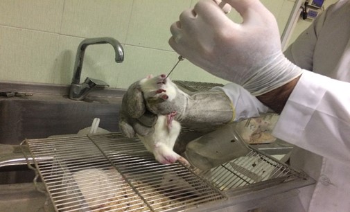
Figure 1. How to gavage extracts in mice
Three hours after the last injection, animals in all groups were killed and their incoming stomach was quickly removed and cut from a large curvature, and then the pathologist examined the type of wound, bleeding, and inflammation. After preparing paraffin sections, the samples were observed with a stereomicroscope (Lica Zoom 2000). Incoming gastric lesions were examined separately and measured using mm using Wild Heerbrugg, Switzerland (Graticule) and their mean was determined. Spotted lesions (petechiae) with a size of one mm were also counted and all five spotted lesions were considered as one mm wounds.
1-2 = 2mm
3-4 = 3mm
6-5 = 4mm
Losses larger than 6 mm were considered equal to 5. The sum of the total amount obtained for wounds was calculated and considered as the wound coefficient (3, 11). The recovery coefficient was calculated from the following formula:
Healing coefficient = 100% Wound control coefficient / Treatment wound coefficient - Wound control coefficient
After microscopic calculations, the stomachs and intestines were placed in the neutral formalin buffer and stabilized and prepared for tissue sections. Six-micron sections were prepared and stained by hematoxylin-eosin method and given to the pathologist for interpretation blindly without knowing the tested groups. Microscopic images were taken with the Olympus CH30 camera and microscope, and macroscopic images were taken directly with the digital camera.
Statistical Analysis
The results were analyzed by one-way Anovoa test using SPSS 11 software and the difference was considered significant with P-value<0.01.
According to the results of Table 1, injection of the aqueous extract of Glycyrrhiza glabra) AEGg) in the mentioned doses did not show any acute toxicity. The results are shown in Table 1.
| The first step of the acute toxicity test | The second stage of the acute toxicity test | ||
| Number of animals / mortality | Dosage and substance | Number of animals / mortality | Dosage and substance |
| 3/0 | AEGg 10 mg/kg | 11/0 | AEGg 1600 mg/kg |
| 3/0 | AEGg 100 mg/kg | 11/0 | AEGg 2900 mg/kg |
| 3/0 | AEGg 1000 mg/kg | 11/0 | AEGg5000 mg/kg |
Rating Test Results
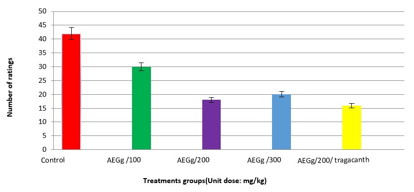
Carrageenan Test Results
| Weight difference between right and left foot | Group |
| 3.79 ±59 | Control |
| 3.50 ±60 | Witness |
| *15/2±11/58 | Root extract (100 mg / kg body weight) |
| ***84/2±50/18 | Root extract (200 mg / kg body weight) |
| ***88/0±32 | Root extract (300 mg / kg body weight) |
| ***73/1±33/10 | Dexamethasone |
Pathological Examination Results
Receiving three consecutive doses of aspirin could cause mucosal lesions in the stomach of mice treated with mucosal lesions and cause bleeding inflammation in the intestine. On macroscopic examination, multiple hemorrhagic lesions of various sizes were observed in the gastric endocrine glands, covering a large portion of the rat gastric gland. On microscopic examination, obvious wounds were seen passing through the mucosal layer and destruction of the epithelial tissue in the entrance stomach. As seen in Figure 3, in the negative control sample of the stomach, the mucosal layer of the gastric mucosa and muscle has a regular and normal appearance and histologically the epithelial and pores are completely healthy. Pathological images of normal rat stomach tissue are shown in Figure 3.
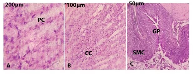
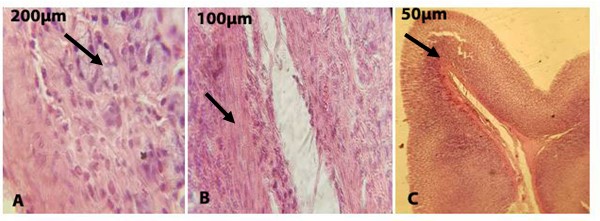
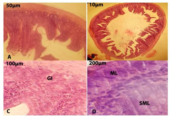
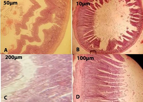
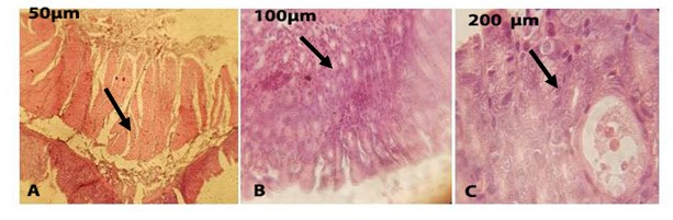
Figure 7. Effect of aqueous extract of G. glabra on gastric ulcer. In Figure (b), the amount of inflammation is reduced compared to the ulcer control sample and the histological structure is completely normal, but a slight exudation of neutrophil cells is observed at the flash.
Figure 8. The effect of aqueous extract of G. glabra on intestinal ulcers is shown. The accumulation of blood cells in the intestinal villi is observed at the flash and the spread of inflammatory cells is observed at the tip of the intestines.
Pathological examinations and calculation of wound coefficient showed that aqueous extract of G. glabra significantly reduced wound coefficient compared to the control group. This lower wound coefficient was also shown in the group receiving tragacanth and aqueous extract of G. glabra (P<0.01).
Also, in the group receiving the aqueous extract of Glycyrrhiza glabra, the wound healing percentage compared to the negative control group (distilled water) was 9.53% and in the group receiving omeprazole was 90.27%. Also, in the group receiving the composition of licorice extract and veterinary, there was no significant difference in wound healing rate compared to the group receiving licorice alone, while it had almost the same effect on increasing the wound healing percentage. (P>0.05) The results of the wound coefficient are consistent with the pathology results. In pathological examinations in the group receiving the aqueous extract of G. glabra in the stomach, gastric mucus was completely normal and mild lymphocyte proliferation and less bleeding were observed than in the control group, which indicates the positive effect of aqueous extract of G. glabra on wound healing. In the positive control group, the wound healing ratio with omeprazole was 69%, which was lower than the groups receiving the aqueous extract of G. glabra and aqueous extract of G. glabra with tragacanth groups. Morphometric evaluation to measure wound spread showed that the wound coefficient in the control groups of distilled water and omeprazole decreased less to the other two treatment groups. The results are shown in Table 3 and Figure 9. In Figure 10, the percentage of wound healing in the treatment groups is compared.
| Groups | Ulcer Index | Recovery ratio compared to omeprazole | Recovery ratio compared to distilled water |
| AEGg | 2.95 | 90.27% | 91.53% |
| AEGg+Tragacanth | 3.2 | 91.85% | 93.12% |
| Omeprazole ( Positive Control) |
20.44 | - | 69% |
| Distilled Water (Negative Control) |
32.38 | - | 0 |
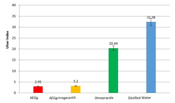
Figure 9. Comparison of wound coefficient in the treatment groups compared with control groups (P<0.01)
Figure 10. Comparison diagram of recovery percentage in the treatment groups compared with control groups (P<0.01)
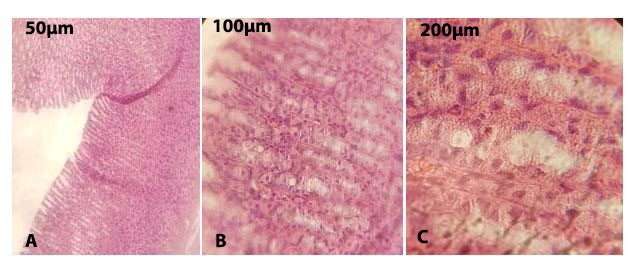
Figure 11. Effect of aqueous extract of G. glabra and tragacanth detoxification of gastric tissue. According to the figure above, the histopathological pattern is completely normalized
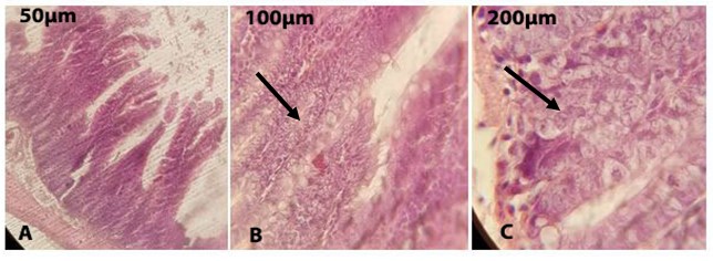
Figure 12. Effect of aqueous extract of G. glabra and veterinary detoxification on intestinal tissue. Mild inflammation and necrosis of a number of lymphocyte (PMN) cells are still seen at the site of Willy swelling after consuming the aqueous extract of G. glabra.
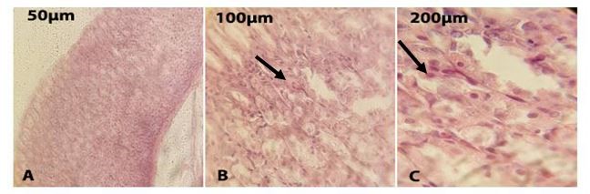
Figure 13. Microscopic picture of pathological ulcer in the stomach in the omeprazole treatment group (Flash: shows a slight lymphocyte infiltration, indicating that the omeprazole treatment does not improve 100%).
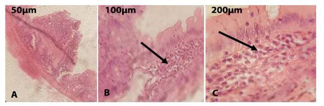
Figure 14. Microscopic picture of pathological ulcer in the intestine in the omeprazole treatment group (flash: shows high neutrophil infiltration with dilation of blood vessels in the intestinal villi, indicating a percentage reduction in improvement as a result of omeprazole treatment).
Aqueous extract of Glycyrrhiza glabra (200 mg / kg) has anti-ulcer protective activity in the stomach and anti-inflammatory properties in the intestine and significantly reduces the wound coefficient compared to the two groups of negative control (distilled water) and positive control (omeprazole) (P <0.01) decreases. It also improves wound healing rate compared to 90.27% positive group and 9.53% negative control group. Diagnosis of acute toxicity of various unknown compounds, including aqueous extract of Glycyrrhiza glabra, was evaluated using LD50. In fact, the LD50 is an important indicator for detecting the minimum allowable dose in order to kill 50% of the animals (23, 25). In the present study, no mortality occurred after 72 hours after injection of different doses of the extract, so it is possible that the aqueous extract of Glycyrrhiza glabra is completely safe and harmless in both stages of acute toxicity testing, at least in doses between 10 and 5000 mg/ kg. Rating test is used to identify environmental mechanisms and acetic acid used in this test can activate endogenous compounds such as bradykinin, serotonin, histamine.
The results showed that the aqueous extract of Glycyrrhiza glabra prevented abdominal contractions caused by acetic acid. Therefore, a possible cause of the anti-inflammatory and anti-infiltration effect of neutrophils, the extract may be due to inhibition of the release of endogenous compounds (arachidonic acid metabolites) and it is speculated that its sedative effects are supported by environmental mechanisms (23). The results of the present study showed that the aqueous extract of licorice root at a dose of 200 mg / kg was able to reduce the inflammation induced by carrageenan and rating tests, whose anti-inflammatory effect was not comparable to dexamethasone. Aqueous extract of Glycyrrhiza glabra contains glycyrrhizin, liquoricidine, saponin, glycyrrhizic acid, a toxic glycyrrhizin, lactic acid, which, according to current sources suggest that a phenolic substance called glycyrrhizin, which is one of the main constituents of licorice, It has a positive effect on inflammation caused by gastric ulcer in rats and has caused its partial treatment (18).
This is probably due to a change in the antioxidant activity of the plant, which has reduced inflammation by inhibiting protein kinase C, phospholipase A and phosphodiesterase, as well as other inflammatory factors such as prostaglandins and histamine (16, 25).
Factors such as gastric ulcers enter the body through mechanisms that increase calcium and free radicals, and in the process leading to the release of arachidonic acid and the enzyme cyclooxygenase, which converts it into a prostaglandin precursor, which in turn causes inflammation. The results of the present study show that the aqueous extract of Glycyrrhiza glabra inhibits inflammation in the rating test and carrageenan, but more research is needed to determine the exact mechanisms of their effect and possible pathways of anti-inflammatory action (23 , 24).
In the study of Jalilzadeh-amin et al. (2015), alcoholic extract of licorice with a dose (50-150 mg/ kg) was able to show the anti-ulcer effects of gastric ulcer, which differs from the dose used in this study. Of course, the results of this study are consistent with the results of the present study regarding the percentage of wound healing, but due to the use of ethanolic extract in Jalilzadeh's research on reducing the inflammatory effects differ from the results of this study, because in this study aqueous extract was used and ethanol effect. As a separate parameter, it could be effective in reducing the rate of inflammation. On the other hand, the dose of 200 mg/ kg was given as peritoneal injection, but in this study, the dose was used as a gavage and was directly related to intestinal and gastric cells. The place has been effective, which of course requires more research (12). In the microbiological studies, there was no significant difference between the results obtained in the group that received an aqueous extract of Glycyrrhiza glabra and tragacanth together with the group that received only aqueous extract of Glycyrrhiza glabra (P> 0.05), but in the pathological studies, this difference was significant. This result indicates that the association of tragacanth with aqueous extract of Glycyrrhiza glabra is not only effective in relieving histopathological symptoms in the stomach and intestines to some extent, but also according to articles can reduce the side effects of licorice consumption such as high blood pressure. Proves the superiority of licorice over existing chemical drugs such as omeprazole. (P <0.01) Of course, the synergistic effect of tragacanth and licorice, using different doses, should be investigated in another study.
In a study by Kjayyal et al. (2001), they studied the effects of aqueous extracts of Lemongrass, Siberian, Chamomile, Cumin, Mint, Licorice, Angelica and Marianlu thistle on the anti-ulcer activity of these extracts in combination in the gastrointestinal tract. The results of antitumor activity of the extracts were confirmed histologically and studies showed that the use of several extracts together based on the dose can reduce the amount of acid and increase mucin secretion, increase the release of prostaglandin E2 and decrease leukotrienes. The effect on pepsin content was relatively variable and did not appear to be related to antitumor activity. The most effective effect of aqueous extract of Glycyrrhiza glabra and asparagus at a dose of 10 mg / kg of rat body compared to 100 mg / kg of cimetidine was shown on the rat gastric ulcer, which differs from the dose of aqueous extract in this study. In this study, a combined aqueous extract of Glycyrrhiza glabra and tragacanth at a dose of 200 mg / kg was used in comparison with omeprazole. The difference between the dose in this study and the above study shows that based on the type of active ingredients in each plant, even with the same efficiency, the dose is different and the synergistic effect of plant extracts is very different based on their dose and even possible side effects. Extracts can be combined with a dose-based manner that requires extensive laboratory research (10).
In the study of Nafeeza et al. (2002), aspirin was very effective in causing ulcers only in the gastric lining and had no effect on intestinal tissue, but However, in this study, according to the results, tissue changes occurred in the intestinal tissue, which was less severe than in the stomach and is quite evident in the pathology images taken. The rate of inflammation has decreased, but the aqueous extract of Glycyrrhiza glabra continues to reduce the wound rate in the gut and increase the healing rate. (P <0.01) The results of this study are different from the present study. (13) The etiology of gastric ulcer is still debated, but there is an imbalance between invasive factors and gastric mucosal defense mechanisms. Many people use NSAIDs daily to relieve digestive problems, especially stomach pain, and recent studies show that between taking this Pill and the occurrence of gastrointestinal lesions are associated with the release of free radicals (14).
Some herbs can have a positive effect on gastrointestinal function. According to some studies, herbs such as Glycyrrhiza glabra by inhibiting acid, increasing mucus production, stabilizing superficial epithelial cells and interfering with the production of prostaglandins in preventing ulcers and They are effective in wound healing (15).
Ramirez et al. (2004) also showed that the extract of some plants that have similar compounds in Glycyrrhiza glabra has an anti-ulcer effect on gastric ulcer, which the results of this study confirm this, of course, the effect of Glycyrrhiza glabra. In the intestine, it is only to the extent of reducing inflammation and complete recovery in intestinal lesions has not been achieved. (16) Glycyrrhiza glabra is a genus of herbaceous plants and due to the active compounds present in the rhizome of this plant, it has known anti-ulcer properties and often has compounds of flavonoids, isoflavones, hydroxycoumarin, alkali derivatives, steroids and Fragile oils such as estragole and hexanoic acid have many fatty properties, including antioxidant properties. In the studies of Nolan et al. (2005) the protective effect of flavonoids on the improvement of wound ulcers in the gastric lining has been shown to be pathologically consistent with the present study (17).
A study by Jia, T et al. (2017) found that another flavonoid called licoriceidin is present in Glycyrrhiza glabra root, which has anti-inflammatory effects on chondrocyte osteoarthritis. It is oxidized to prostaglandins and nitric oxide, followed by the Nrf2 pathway, a lotion-based protein and transcription factor encoded in humans by the NFE2L2 gene, by inhibiting oxidative compounds from free electron production and increasing extracellular calcium, thereby causing inflammation. Prevent. Liquoridine is an antispasmodic and anti-inflammatory, antioxidant found in Glycyrrhiza glabra root (20).
The results of this study had an inhibitory effect on chondrocyte inflammation and in this study the use of aqueous extract of Glycyrrhiza glabra in the stomach reduced lymphocyte diffusion and bleeding compared to the control group which indicates the positive effect of aqueous extract of Glycyrrhiza glabra on wound healing. The use of aqueous extract of Glycyrrhiza glabra and tragacanth completely erases the pathological changes and shows a completely normal histopathological pattern, which clearly shows the synergistic effect of aqueous extract of Glycyrrhiza glabra and tragacanth compared to the results of Jia T research (20).
Although the anti-ulcer effect of aqueous extract of Glycyrrhiza glabra can be attributed to the chemical compounds in the plant extract, its exact mechanism remains unknown due to the few studies available. Increasing free radicals as a result of aspirin consumption and the effect of vitamin C in reducing them and the protective effect of flavonoids, sterol and isoflavones by strengthening the mucosal barrier are some of the things that protect the plant against aspirin ulcers in the stomach and reduce aspirin-induced inflammation in the intestine. The cheapness, nativeness and availability of these plants are important factors that lead researchers to use medicinal plants in the treatment of infections. The results of the present study show that the aqueous extract of Glycyrrhiza glabra is effectiveness of healing of gastric ulcer and the relative effect on the healing of intestinal ulcers. The synergy of tragacanth and Glycyrrhiza glabra completely eliminates the pathological changes and creates a normal histopathological pattern in the stomach and a relatively normal one in the intestine. In future studies, the possible pathways of anti-inflammatory action should be identified by accurately identifying the active ingredient of Glycyrrhiza glabra and tragacanth and determining the exact mechanisms that improve their effect in the treatment of gastrointestinal infections. According to the results of this study and the mentioned properties of Glycyrrhiza glabra and the absence of side effects on the body, the use of Glycyrrhiza glabra and tragacanth is recommended based on the instructions of experts.
This research is one of the research projects of the Sampad High School in Tehran, which has been carried out in the laboratory of the Islamic Azad University, North Tehran Branch and the boarding house of Baqiyatallah University. The authors thank Dr. Barkhordari and Dr. Amini.
Funding for this research has been entirely the responsibility of the authors.
Authors declared no conflict of interests.
Received: 2020/03/12 | Accepted: 2020/04/25 | ePublished: 2020/07/20
| Rights and permissions | |
 |
This work is licensed under a Creative Commons Attribution-NonCommercial 4.0 International License. |
Copyright Policy
Iranian Journal of Medical Microbiology by Farname is licensed under CC BY-NC 4.0![]()
![]()
![]()
