


![]()
![]()
![]()
BibTeX | RIS | EndNote | Medlars | ProCite | Reference Manager | RefWorks
Send citation to:
URL: http://ijmm.ir/article-1-1249-en.html


2- Department of Pathobiology, Faculty of Veterinary Medicine, Shahrekord University, Shahrekord, Iran. ,
3- Department of Pathobiology, School of Veterinary Medicine, Shiraz University, Shiraz, Iran.
.
Canine distemper virus (CDV) is a member of the family Paramyxoviridae, which can infect certain epithelial cell lines of the respiratory, gastrointestinal, or nervous systems (1). The virus is endemic in many areas, such as India, Denmark, Finland, Brazil, and North America (2). A high prevalence (55.6%) of CDV has been reported among the rural dogs in the north of Iran (3). Avizeh et al. (2007) in another study revealed a prevalence of 17.52% for CDV (4).
In sensitive species, CDV inhibits immunity and its receptor is the signaling lymphocyte activation molecule 4 (SLAM 4) (5). Such condition often gives other microorganisms a chance of occurrence and infection. Although the distemper vaccine has significantly reduced the disease, it is still endemic in many parts of the world (6-12).
There are studies on the co-infection of CDV and some other viruses, including canine parvovirus type 2 (CPV-2), canine alphaherpesvirus 1 (CHV-1), and canine adenovirus types 1 and 2 (CAV-1 and -2). Recently, new reports have shown that CDV infection can facilitate the development of diseases caused by emerging viruses (13-17).
Morbilliviruses, such as CDV are transmitted by aerosols and cause clinical signs, including respiratory and gastrointestinal symptoms, which are often complicated by other pathogens. Another feature of Morbillivirus infection is the temporary severe suppression of the host immune system (7).
During the first 24 h after CDV infection, the virus accumulates in macrophages, as well as B and T cells. Afterwards, the virions go towards the lymphoid tissues and multiply in the reticuloendothelial organs within 4-6 days after the infection (18, 19). The initial virus proliferation in the lymphoid tissues leads to severe suppression of the immune system (20, 21, 22).
Following viraemia, CDV spreads to a number of epithelial tissues and the central nervous system (19). Infectious lymphocytes penetrate the epithelial tissue and may locally increase the number of viruses in the epithelial cells, followed by respiratory, intestinal, and urinary tract infections. Penetration of T lymphocytes and dendritic cells can, in turn, cause cutaneous disease manifestations (23). Moreover, lymphopenia could occur due to a reduction in the population of CD4+ and CD8+ T cells, in addition to CD21+ B cells (21, 22). Unlike CD8+ cells, which are resistant, CD4+ lymphocytes, as apoptotic cells, are likely to die (23, 24, 25).
The decline in circulating immune system cells may result from the diminished number of cells in the lymphatic organs and the apoptosis of white blood cells. Furthermore, programmed cell death can happen in non-infected cells indicating an additional non-viral mechanism of apoptosis. Therefore, the overactivation and death of lymphocytes because of the Fas protein should be considered in the CDV infection (26, 27). On the other hand, canine parainfluenza virus type 2 (CPiV-2) is an ssRNA virus belonging to the Paramyxoviridae family. This virus is quite similar to the simian virus 5, which was first reported as Simian-5 virus by Binn et al. in 1967 in dogs with respiratory disease symptoms.
The CPiV-2 is a highly contagious virus, an important symptom of which is a dry cough. Mild fever, lack of energy, and loss of appetite are other symptoms of the disease. Collecting and keeping dogs in shelters and breeding centers or contact with sick dogs can cause further spread of the disease.
Vaccine administration in puppies can protect dogs from the virus. Puppies are more vulnerable to pneumonia than adult dogs. Infection usually leads to short-term coughs along with a number of general symptoms. Despite vaccination, the virus is still one of the most common respiratory pathogens of dogs (28). With this background in mind, the present study aimed to perform the molecular and immunological investigation of CDV and coinfection of this virus with CPiV-2.
Sampling
Samples were taken from dogs in the clinics of Isfahan (2018-2019). The age of the dogs was determined based on the statements of their owners and the examination of the animal's teeth. For sampling, the animals were first physically restrained and examined for clinical signs. Following physical restraint and sometimes using ketamine (Alfasan, Netherlands) and acepromazine (Hoogstraten, Belgium), respiratory and gastrointestinal tract swabs were taken from 50 symptomatic and 50 asymptomatic dogs. The specimens were examined by a rapid immunochromatography detection kit of distemper and were placed in sterile phosphate buffer saline.
Rapid Distemper Immunochromatography
A rapid immunochromatography kit of distemper (VetALL SensPERT Canine Distemper Virus Test Kit, CDV Ag) was applied for the detection of CDV in the eye and respiratory swabs. The samples were first diluted in the kit buffer. Next, 100 µL of the reagent was poured into the sample well and time was recorded. Next, the sample was dripped on a pad and after the adsorption of dripped samples by the pad, two red bands showing the positive result appeared on the C (control) and T (test) positions.
Acid Nucleic Extraction and Reverse Transcriptase Reaction
QIAamp Viral RNA Mini Kit (QIAGEN, Germany, Cat No: 52904) was used for RNA extraction from buffy coat. To determine the quality of the extracted RNA samples, the optical density of the specimens was measured at 260/280 nm by a spectrophotometer. Moreover, cDNA synthesis was carried out utilizing TaqMan reverse transcriptase (RT) kit (Invitrogen, USA, Cat No: N8080234). The RT reaction entailed 1 μL of 10X RT buffer, 2.2 μL of 25 mM MgCl2, 2 μL of 10 mM dNTPs, 0.5 μL of random hexamers, 0.2 μL of RNase inhibitors, 0.25 μL of M-MLV RT (50 U/μL), RNA template (400–500 ng/μL), and deionized sterile water up to 10 μL. Moreover, the thermal cycling program encompassed 25oC for 10 min, 48oC for 30 min, and 95oC for 5 min.
RT-Polymerase Chain Reaction and Statistical Analysis
Polymerase chain reaction (PCR) was conducted using primers designed by Beacon Designer software for the detection of CDV and CPiV-2. The sequences of primers were as follows: CadF: AAGCCTCACACTGTTCAAG -3’, CadR: GATTAGGACTATAATGACATGC -3’, and Capi2F: ACAATCCCACCTACAACAC -3’, Capi2R: 5’- AATCCGTAGGCAGAATAG -3’.
For detecting CPiV-2, the PCR thermal cycle programs consisted of 5 min at 94ºC, 30 cycles of 45 s at 94ºC, 45 s at 59ºC, 45 s at 72ºC, and 5 min at 72ºC. For CDV, the PCR cycles and temperature were the same as CPiV2. The only difference was that the annealing temperature was 57ºC. In each test, the negative and positive controls prepared from Tehran University were also applied.
The PCR products were electrophoresed in 1.5% agarose with 0.01% Green viewer (Parstous, Iran, Cat No.: B111151) along with 50 base-pair ladders (Fermentas, USA, Cat No.: SM1133). Afterwards, two positive PCR products were sent for sequencing to Bioneer, Korea. Finally, the data were analyzed using the Chi-square test by the SPSS 16 (IBM, Ill., USA) version 16.
There were 29 and one positive cases among symptomatic and asymptomatic dogs based on the results of rapid distemper immunochromatography kits, respectively. The CDV-positive samples and positive control had a band size of 100 bp, while CPiV-positive specimens and positive controls had bands of 102 bp in size (Figures 1 and 2). Moreover, the sequencing of RT-PCR products showed that the degree of the identity of the sequences with the sequences registered in the GenBank for CDV and CPiV-2 was high. This was a good indicator of PCR efficiency (Figures 3 and 4).
In general, we detected 37 and three CDV-positive cases using RT-PCR among 50 symptomatic and 50 asymptomatic dogs, respectively. In addition, the frequency of CPiV-2 cases in the first and second groups was 11 and one cases, respectively. The rate of CDV and CPiV-2 coinfection was 4% only among dogs with distemper clinical signs.
All the 30 positive samples detected by rapid distemper immunochromatography test were also positive by RT-PCR test. Furthermore, there was a statistically significant relationship between the results of these two tests (P<0.05). However, we noticed no statistically significant correlation between the two studied diseases and gender (P>0.05). However, the relationships of CPiV RT-PCR results and CDV signs with age were statistically significant (P<0.05). The relationships of CDV RT-PCR results and CDV signs with age were statistically significant (P<0.05)

Figure 1. Electrophoresis gel of CDV RT-PCR products.
1: Negative control, 2,8: CDV positive RT-PCR products (100 bp), 6: Positive control (100 bp) 6: Ladder (50 bp)
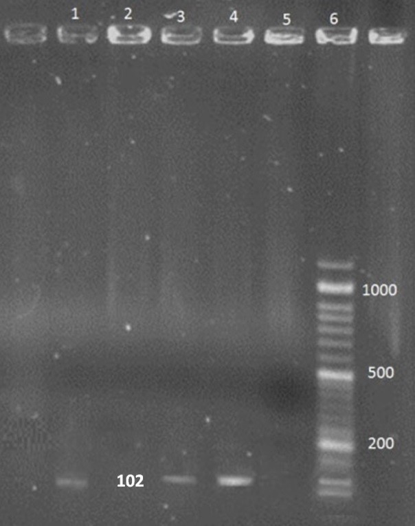
Figure 2. Electrophoresis gel of CpiV-2 RT-PCR product.
1,3: CpiV-2 (102 bp) positive PCR product,2: Negative control, 4: Positive control, 6: Ladder (50 bp)

Figure 3. Alignment of CDV RT-PCR product sequence with the sequences registered in the gene bank.

Figure 4. Alignment of CpiV-2 RT-PCR product sequence with the sequences registered in the gene bank
Almost all dogs with distemper experience immune suppression. As a result, other infections can be added to the disease and make these dogs sicker. The mechanism of immune suppression in distemper disease is not fully understood. This mechanism depends on several factors. If some white blood cells become infected, long-term immune disorders might continue even after the peripheral blood is cleared of the virus during the recovery period (5).
Therefore, immune-suppressing mechanisms may affect the cells that are not directly involved in viral infection. Although the exact mechanism of immunosuppression due to CDV infection is not yet well understood, the immune system weakness due to this virus is accepted by all scientists.
Symptoms of the early stage of distemper in dogs usually appear as eye and nose discharge, fever, loss of appetite during which other canine respiratory infections may occur. Consequently, it is impossible to distinguish the infections exclusively based on the symptoms. Therefore, considering the possibility of concomitant infections due to immunosuppression resulting from CDV, the current study was conducted to investigate coinfection with distemper and canine parainfluenza type 2. We found coinfection with these two viruses in 4% of the cases with no statistically significant relationship between distemper and canine parainfluenza virus-2.
In the present study, unfortunately, limited access to dogs with distemper clinical symptoms did not allow us to have a large sample size and achieve a significant relationship between the two studied viruses. A rapid distemper diagnostic kit is the best option for diagnosing the virus in veterinary clinics. The specificity and sensitivity of this kit for conjunctival specimens, similar to Nested PCR, is 100%. (29). There is not any interference between vaccination and this diagnostic method. Therefore, this assay can be easily used in areas without diagnostic facilities (18, 28). All the above-mentioned reasons led us to choose this technique for the initial diagnosis of CDV.
Previously, some research groups worked on CDV coinfection with other pathogens. In Iran, we could not find any reports of infections simultaneous with distemper. There were only some reports of CDV infection from some cities. For instance, Avizeh et al. in 2007 carried out a serologic detection of CDV in unvaccinated dogs from Ahvaz, Iran. Furthermore, Namroudi et al. (2015) detected the Arctic and European clusters of CDV in the north and center of Iran (3, 4).
Moreover, we found some related works in other countries. For example, in Japan, Mochizuki et al. (2008) investigated the pathogens involved in upper respiratory tract infections in domestic dogs. In the present study, CPiV (4.7%), canine coronavirus 1 (4.4%), CAdV-2 (2.9%), canine coronavirus 2 (1.5%), and CDV (1.5%) were detected and two cases of coinfections were observed (14).
In a research performed by Damián et al. (2005), CDV, CAV, and CpiV were detected in 77%, 57%, and 51% of the studied population, respectively. The most common coinfection was found to be CDV-CpiV in 14% of the cases (20). Headley et al. (2018) reported CDV-associated infections as Neospora caninum (100%), CPV-2 (100%), CAdV-1 (100%), and CAdV-2 (100%) (11).
Aguiar et al. (2012) assessed the coinfection of CDV and Toxoplasma gondii in dogs with neurological signs. Using RT-PCR, 80.9%, 38.1%, and 41.1% of dogs were positive for CDV, anti-T. gondii antibodies, and both factors, respectively (3). Furthermore, Headley et al. in 2015 detected the coinfection of CDV with CHV-1, CAdV, and CPV in domestic dogs in southern Brazil (11).
The mentioned research indicated a high rate of CDV coinfection with other pathogenic viruses in dogs. This rate confirms the hypothesis that susceptibility to other pathogens due to the weakness of the animal immune system elevates after CDV infection. However, performing further similar studies on other virus hosts can give us more comprehensive information about this issue.
The high prevalence of distemper, the possibility of concurrent infections with CDV in Iran and other endemic areas, and the reports of distemper from other hosts suggest that distemper and simultaneous infections in wildlife, rodents, and other species associated with CDV infections need to be investigated.
In the present research, out of the samples taken from symptomatic dogs, a total of four cases were detected to be co-infected with CDV and CPiV-2. Furthermore, no statistically significant correlation was observed between distemper and CPiV. For future research, we recommend similar studies with an appropriate sample size to obtain more accurate results.
This work was supported by grants from Shahrekord University (Grant number: 98GRD30M1801).
The authors report no conflict of interest.
Received: 2020/11/17 | Accepted: 2021/02/13 | ePublished: 2021/04/9
| Rights and permissions | |
 |
This work is licensed under a Creative Commons Attribution-NonCommercial 4.0 International License. |
Copyright Policy
Iranian Journal of Medical Microbiology by Farname is licensed under CC BY-NC 4.0![]()
![]()
![]()
